COMPANY
INSIGHT
Sponsored by

Unleashing the full potential of 3D orthopedic diagnostics
Weight-bearing CT imaging has introduced new aspects to the diagnosis of foot and ankle diseases. Since its launch in 2012, Planmed’s Verity CBCT scanner has been used in challenging imaging cases of the upper and lower extremities. Now the 3D imaging capabilities are combined with innovative automatic 3D analysis of the image with Disior InGrid™ sofware.
Planmed has joined forces with a Finnish start-up company Disior. This collaboration will transform the way orthopedic imaging and diagnostics are carried out by combining the 3D innovations of two Finnish companies.
Cone beam computed tomography (CBCT) is a technology in which a series of low-intensity 2D X-rays are captured with a flat panel detector. The image series captured during a single or partial rotation is next reconstructed to a 3D volumetric image of the region of interest. This step is achieved using a combination of FDK and iterative algorithms.
CBCT is currently the only technology which is able to produce 3D volumetric imaging data of the foot and ankle under real, natural weight-bearing conditions. Combining the weight-bearing imaging with the fast image acquisition, low patient radiation dose and isotropic spatial resolution of 0.2mm allows the client to maintain complete confidence in their diagnostic diagnosis.

The Disior InGrid algorithm effectively solves the problems related to 3D measurements of bones and joints. We are very excited about this says Juhamatti Malm of Planmed
Taken to 3rd dimension
Conventional 2D weight-bearing X-ray imaging has long been used as the standard of care to visualize skeletal diseases, and deformities of the foot and ankle. The measurement techniques are well described in the literature. However, 2D data is prone to projection differences and the superimpositions of the bones on one another can mask underlying issues.
3D weight-bearing imaging solves the challenges of projection differences and overlapping structures. The anatomy is shown in a naturally occurring position and can better reveal joint space narrowing and other conditions that may not be visible through conventional diagnostic means.
Adding the third dimension allows the anatomy to be visualized as 2D transversal slices. 2D transverse views enable the manual measurement of foot and ankle diseases and deformities. However, performing accurate and repeatable 3D measurements is not as easy as one might think.
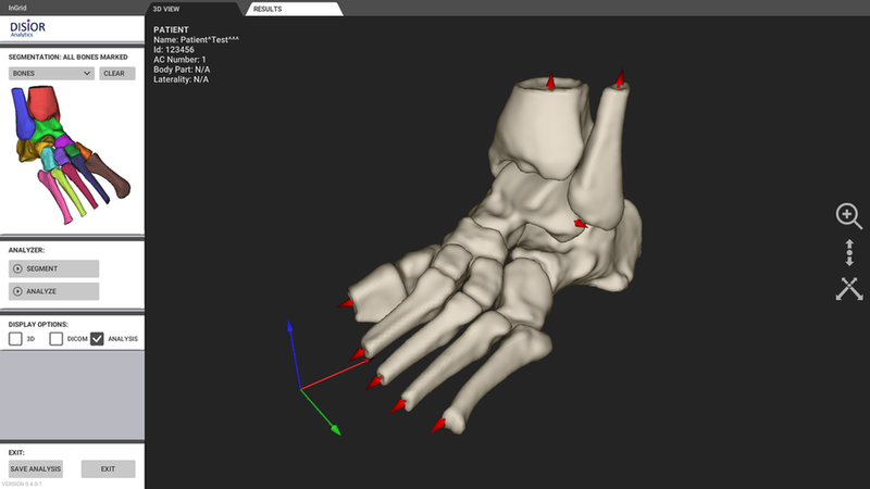
The weight bearing 3D image acquisition and an example of Disior InGrid processing analysis on the patient’s foot.
A learning curve
Additionally, more complex midfoot and hindfoot alignments and commonly used Saltzman view are easily available from the 3D weight-bearing data.
Combining high quality 3D weight-bearing images taken with Planmed Verity® with clever and automatic 3D measurements has key implications for diagnosing foot and ankle anatomy. It carries great potential in clinical decision-making, surgical planning and post-operative analysis. In addition the outcome of the Disior InGrid™ processing has excellent quality for 3D printing of anatomic models.
Removing the chance of human error
The Disior InGrid algorithm solves the problems related to manual 3D measurements of bones and joints. It uses deformable shape models to segment bone tissue from imaging data.
Using the 3D image captured with Planmed Verity, the algorithm automatically extracts the landmarks and longitudinal axes needed for the measurements from the segmentation result using robust feature detection algorithms. This results in excellent measurement repeatability, without the steps prone to human error.
Transforming a DICOM image into a mathematical model of the anatomy serves as the basis for an analysis of the anatomy and kinematics. Based on this analysis, any 3D measurement can be taught and automatised in the software. As a result, clinicians and radiologists are no longer dependent on individual slices, but may easily use the whole data set for analysis. This way, transferring the measurement and diagnosispractices from weight-bearing 2D to 3D is easily achieved.
With the Planmed Verity and Disior InGrid algorithm, one can easily add value to the diagnosis of common clinical cases in foot and ankle region.
For example, hallux valgus, flat foot and cavus foot are analysed in an unseen way by automatically measuring angle, such as the first and second intermetatarsal angle, or Meary’s angle, from 3D weight-bearing data. Also more challenging analyses such as joint space distance mapping are now possible.
Straightening of the wire is one of our core competencies.
Further information
Juhamatti Malm
Director, Product Portfolio and Business Development

CONTACT INFORMATION
Planmed
Sorvaajankatu 7
00880 HELSINKI
FINLAND
Tel. +358 20 7795 300 (switchboard)
Fax +358 20 7795 664
Fax: +49 (0) 6172 27978-10

next article